Successful Sphincter-Preserving Excision of Giant Gangrenous Grade IV Haemorrhoids Using LigaSure Vessel Sealer with a Novel Intraoperative Index-Finger Protection Technique
Senior Consultant Surgeon, Bhandari Piles Hospital, Chittorgarh, Rajasthan, India
Honorary Consultant Surgeon, M.P. Birla Hospital and Research Centre, Chittorgarh, Rajasthan, India
Correspondence: opbhandari@hotmail.com · +91-9414109124
Abstract
Background: Prolapsed haemorrhoids (Grade IV) represent the most severe form of anorectal disease, posing extraordinary surgical challenges. When the haemorrhoidal mass approaches dimensions of twelve centimetres, the risk of faecal incontinence from inadvertent damage to the anal sphincter complex is severe.
Case Report: A patient in his early fifties presented with Grade IV haemorrhoids of approximately 12 cm — comparable in size to his thigh — irreducible for five years, with a 15-year history of prolapse. Surgical excision was performed at Dr. Bhandari Piles Hospital, Chittorgarh, using a LigaSure Advanced™ vessel-sealing device. A novel intraoperative technique was employed: continuous placement of the non-dominant index finger inside the anal canal throughout excision, providing real-time tactile identification and physical protection of the sphincter musculature. Post-operative wound management utilised twice-daily autoclaved magnesium sulphate (MgSO4) gauze dressings.
Conclusion: Even extraordinarily large, gangrenous prolapsed haemorrhoids can be safely excised with complete sphincter preservation. The index-finger protection technique, combined with LigaSure technology and structured MgSO4 dressing, offers a reproducible, sphincter-saving approach with full recovery achievable within two months.
Keywords: haemorrhoids; prolapsed piles; sphincter preservation; LigaSure vessel sealer; Grade IV haemorrhoids; haemorrhoidectomy; magnesium sulphate dressing; anal sphincter protection; index-finger technique; gangrenous haemorrhoids
Introduction
Haemorrhoidal disease is one of the most prevalent anorectal conditions worldwide. The Goligher classification grades haemorrhoids I through IV, with Grade IV representing irreducible prolapse — the most severe and surgically challenging presentation.1 Truly giant prolapsed haemorrhoids whose combined mass approximates the size of a major body part are exceedingly rare and appear only in isolated case reports from specialised proctology centres.
The principal surgical risk in such cases is injury to the internal and external anal sphincter complex, which can result in faecal incontinence — a devastating and largely irreversible complication profoundly affecting quality of life. Standard haemorrhoidectomy techniques (Milligan-Morgan, Ferguson) are challenging when tissue volume obscures the operative field and sphincter landmarks.2
We describe a case of 4th-degree gangrenous haemorrhoids with a mass of approximately 12 cm — comparable to the patient’s thigh — managed by a novel intraoperative sphincter-protection manoeuvre using continuous index-finger placement, combined with LigaSure™ vessel-sealing technology. This case has not been previously reported or published in any journal.
Case Report
Patient Details
| Age / Gender | Early fifties / Male |
|---|---|
| Date of Operation | April 2021 |
| Date of Discharge | Approximately one month post-operatively |
| Operated By | Dr. O. P. Bhandari, MBBS; MS (General Surgery); FAIS |
| Personal History | Vegetarian, non-smoker, non-alcoholic |
| Family History | Positive (both parents) |
Chief Complaints
Prolapse of haemorrhoids for five years, irreducible for the last five years. Bleeding per rectum: 15 years of intermittent haemorrhoidal bleeding. History of injection sclerotherapy for piles 10 years prior, performed by another clinician. Massive external prolapse approximating the size of the patient’s thigh (~12 cm mass), consistent with 4th-degree gangrenous haemorrhoidal disease. Inability to reduce the prolapse manually. Significant discomfort, difficulty with ambulation and perianal hygiene.
Clinical Diagnosis
Preliminary diagnosis: 4th-degree thrombosed gangrenous haemorrhoids. Final diagnosis: 4th-degree gangrenous haemorrhoids with irreducible massive prolapse (~12 cm, size of the patient’s thigh).
Physical Examination Findings
General: patient was ambulatory but in moderate distress due to the haemorrhoidal mass. Conjunctiva: pink. Sclera: white. Tongue: moist, pink. Lymphadenopathy: absent. Hepatomegaly/splenomegaly: absent. Blood pressure: 110/70 mmHg. SpO2: 98%. No history of asthma, drug reactions, artificial dentures, previous abdominal surgery, or steroid use.
Anorectal examination: massive prolapsed haemorrhoidal tissue circumferentially protruding from the anal canal. The combined haemorrhoidal mass measured approximately 12 cm — comparable in dimension to the patient’s thigh. Tissue was congested, oedematous, and gangrenous in areas, with purple-grey discolouration indicating compromised vascularity. The anal sphincter was intact on digital rectal examination, though its assessment was severely impeded by the mass volume.
Pre-Operative Preparation
Standard pre-operative workup was performed, including complete blood count, renal function tests, blood glucose, coagulation profile, ECG, and chest radiograph. Bowel preparation was performed the night prior. The patient received prophylactic intravenous broad-spectrum antibiotics immediately before the procedure. Spinal anaesthesia was administered in the sitting position.
Operative Technique
Instrument and Positioning
Instrument used: LigaSure Advanced™ vessel-sealing system. This bipolar electrothermal device achieves simultaneous vessel sealing and tissue transection with minimal thermal spread (≤2 mm), making it ideal for haemorrhoidal tissue in immediate proximity to the sphincter musculature.4,5 The patient was positioned in the lithotomy position with adequate perineal exposure.
Surgical Steps
Inspection and assessment of haemorrhoidal extent and vascular pedicles. Gentle attempt at reduction of prolapsed tissue to delineate the anal verge anatomy.
Key — novel sphincter-protection manoeuvre: Throughout the entire excision, the operating surgeon maintained continuous placement of the non-dominant index finger inside the anal canal. This served a dual purpose: (a) as a tactile landmark to continuously identify the internal sphincter and ensure the LigaSure jaws remained superficial to it at all times; (b) as a physical barrier preventing inadvertent deep biting or thermal spread into the sphincter complex.
LigaSure activation and sequential excision of haemorrhoidal bundles at primary pedicles (right anterior, right posterior, left lateral positions), progressing to secondary and satellite masses. Individual ligation and transection of each pedicle; haemostasis confirmed at each pedicle before proceeding. Skin bridges carefully preserved between excision sites to prevent circumferential stenosis. Final inspection confirmed haemostasis, intact sphincter musculature (confirmed by palpation), and satisfactory excision of all haemorrhoidal tissue. No anal packing was placed; the wound was left partially open for drainage.
Post-Operative Management
Wound Dressing Protocol — Autoclaved Magnesium Sulphate Gauze
A structured twice-daily (BID) dressing regimen was initiated from post-operative Day 1 and continued until wound healing was confirmed. Magnesium sulphate (MgSO4) paste was applied to sterile gauze, freshly prepared each day. Each batch was autoclaved daily to ensure sterility. The autoclaved gauze was gently applied to the post-excision wound bed twice daily (morning and evening) following wound irrigation with normal saline. MgSO4 gauze acts as a hyperosmotic dressing: it draws oedema fluid from the wound, reduces swelling, promotes granulation tissue formation, and provides a bacteriostatic environment. Daily autoclaving ensured zero risk of secondary wound infection.
Systemic Post-Operative Medications
Intravenous antibiotics (broad-spectrum) transitioned to oral antibiotics for 7 days. Oral analgesics: NSAIDs and paracetamol on scheduled dosing. Stool softeners and dietary fibre supplementation. Adequate hydration and daily wound inspection.
Follow-Up and Outcome
The patient was reviewed at 1 week, 2 weeks, 4 weeks, 6 weeks, and 2 months post-operatively. At one month, clinical photographs documented excellent wound healing with significant reduction in perianal swelling and healthy granulation tissue. At two months, complete recovery was confirmed: all wounds healed, near-normal perianal appearance, and — critically — full control of anal sphincters. The patient reported no faecal urgency, no incontinence of flatus or stool, and a normal defaecation pattern. Quality of life was dramatically improved.
Clinical Photographs
Photographic documentation was obtained at two time points by the operating surgeon at Dr. Bhandari Piles Hospital, Chittorgarh. Patient identity has been masked in accordance with journal ethics guidelines.
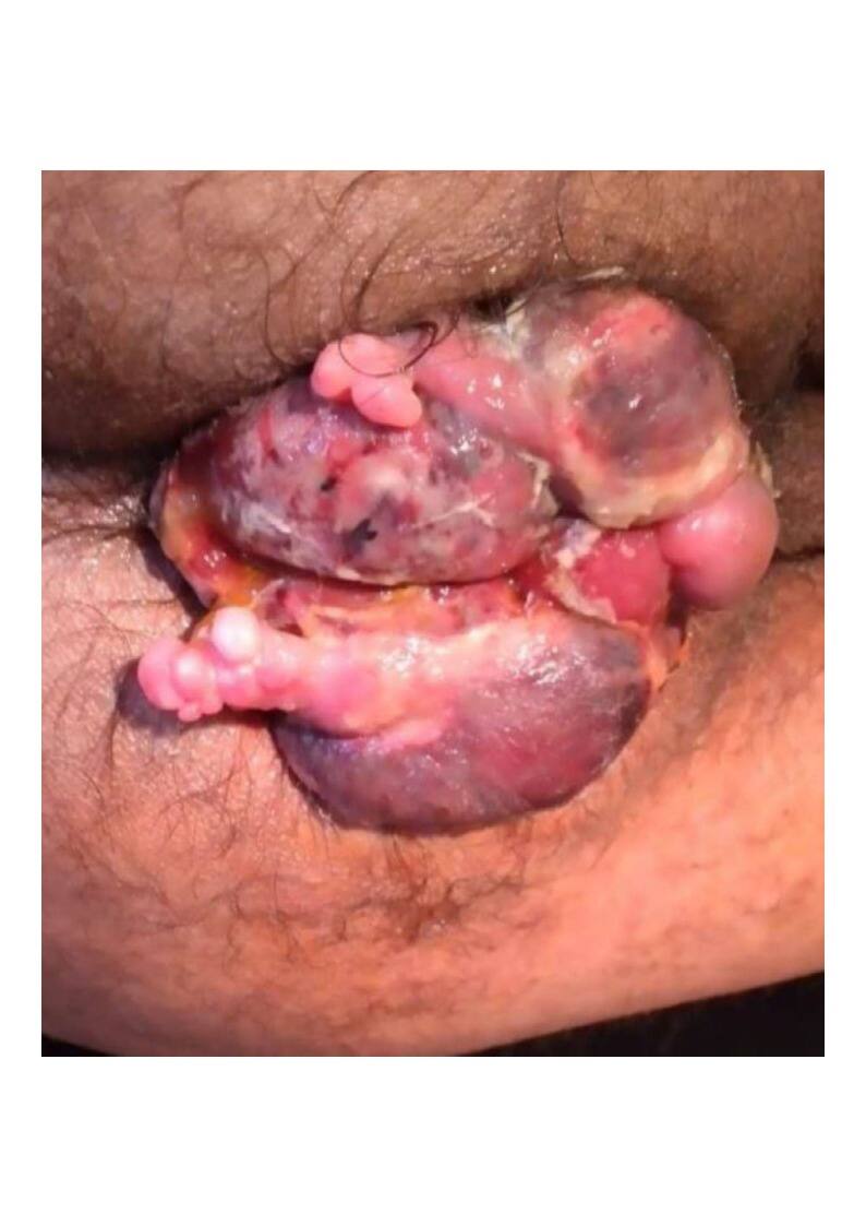
Fig. 1. Pre-operative photograph: massive circumferential prolapsed haemorrhoidal mass (approximately 12 cm; size of patient’s thigh) with gangrenous change (purple-grey discolouration), severe oedema, and tissue congestion.
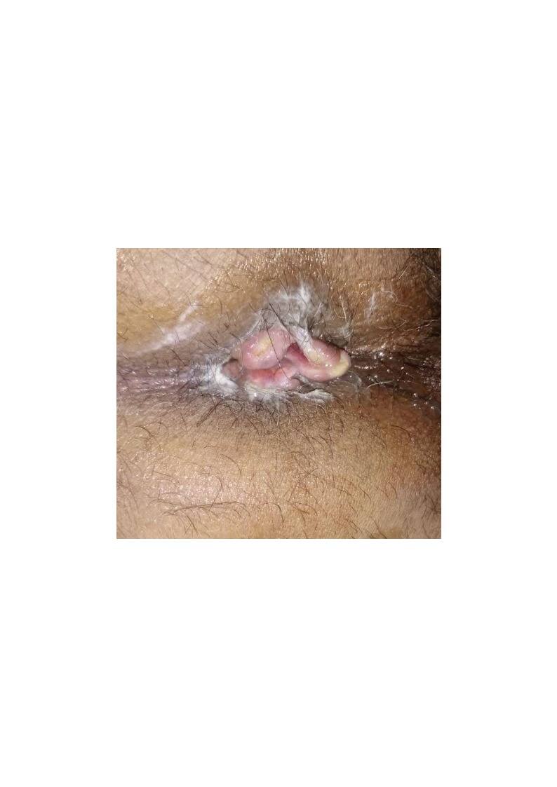
Fig. 2. Post-operative photograph at two months: near-complete wound healing with normal perianal topography restored. Healthy granulation tissue visible. Full anal sphincter control was confirmed clinically.
Discussion
Prolapsed haemorrhoids presenting as a mass comparable to the patient’s thigh are extraordinarily rare and pose unique challenges. A literature review reveals only isolated case reports describing comparable haemorrhoidal mass sizes.6,7 The principal surgical concern — preservation of the anal sphincter — is magnified in such cases because the sheer tissue volume obscures normal anatomical landmarks.
The LigaSure vessel-sealing system has been validated in multiple randomised trials as producing less post-operative pain, less bleeding, and shorter operative time compared to conventional suture-ligation or diathermy haemorrhoidectomy.4,5 Its limited thermal spread (≤2 mm) is particularly advantageous when operating in close proximity to the internal sphincter.
The index-finger protection technique described in this case represents an intuitive but underreported intraoperative safeguard. By maintaining tactile feedback continuously throughout the dissection, the surgeon can identify the sphincter musculature in real time even when overlying haemorrhoidal tissue distorts external anatomical landmarks. This simple yet effective manoeuvre appears to have been the critical determinant enabling complete excision of this extraordinary haemorrhoidal mass without sphincter injury.
The autoclaved MgSO4 dressing protocol addresses the post-operative challenge of managing large wound beds in contaminated perianal environments. The hyperosmotic properties of MgSO4 reduce oedema, promote healing, and prevent secondary infection.8 Daily autoclave cycling of the gauze represents a cost-effective, reproducible approach particularly suitable for resource-limited settings in India.
Conclusion
This case establishes that even giant Grade IV haemorrhoids of extraordinary dimensions — approximately 12 cm in size with gangrenous change and five years of irreducibility — can be safely excised with complete preservation of anal sphincter function. The novel continuous index-finger intraoperative sphincter-protection technique, combined with LigaSure Advanced™ vessel-sealing technology and a structured BID autoclaved MgSO4 dressing regimen, achieved full clinical recovery within two months. This case has not been published in any journal. It is submitted as an original contribution and invites further documentation of the index-finger protection technique in similar cases.
Declarations
- Ethical Approval
- This case report was conducted in accordance with institutional ethical standards. No experimental procedures were performed beyond standard surgical care.
- Informed Consent
- Written informed consent was obtained from the patient for surgical management, photography, and publication of this case report. All patient data have been handled in accordance with applicable privacy regulations. Patient identity has been masked.
- Conflict of Interest
- The author declares no conflict of interest.
- Funding
- No funding was received for this work.
- Author Contributions
- Dr. Om Prakash Bhandari: concept, surgical procedure, post-operative management, data collection, manuscript writing, and final approval.
References
- Goligher JC. Surgery of the anus, rectum and colon. 5th ed. London: Bailliere Tindall; 1984.
- Milligan ET, Morgan CN, Jones LE, Officer R. Surgical anatomy of the anal canal and the operative treatment of haemorrhoids. Lancet. 1937;2:1119–1124.
- Parks AG. De haemorrhoids: a study in surgical history. Guys Hosp Rep. 1955;104:135.
- Tan KY, Zin T, Eu KW, Ong KH, Samuel M, Tang CL. Randomized clinical trial comparing LigaSure haemorrhoidectomy with open diathermy haemorrhoidectomy. Tech Coloproctol. 2008;12(2):93–97.
- Nienhuijs S, de Hingh I. Conventional versus LigaSure haemorrhoidectomy for patients with symptomatic haemorrhoids. Cochrane Database Syst Rev. 2009;1:CD006761.
- Chung CC, Ha JP, Tai YP, Tsang WW, Li MK. Double-blind, randomized trial comparing Harmonic Scalpel hemorrhoidectomy, bipolar scissors hemorrhoidectomy, and scissors excision: ligation technique. Dis Colon Rectum. 2002;45(6):789–794.
- Eu KW, Seow-Choen F. Functional problems in adult rectal prolapse and controversies in surgical treatment. Br J Surg. 1997;84(7):904–911.
- Ozturk E, Yilmazlar T. The importance of wound care in anorectal surgery: a review. Tech Coloproctol. 2010;14(Suppl 1):S17–S21.