“Necessity is not merely the mother of invention. In a resource-limited surgical setting, necessity — and only necessity — is also the teacher, the supervisor, and the examiner.”
I. Introduction
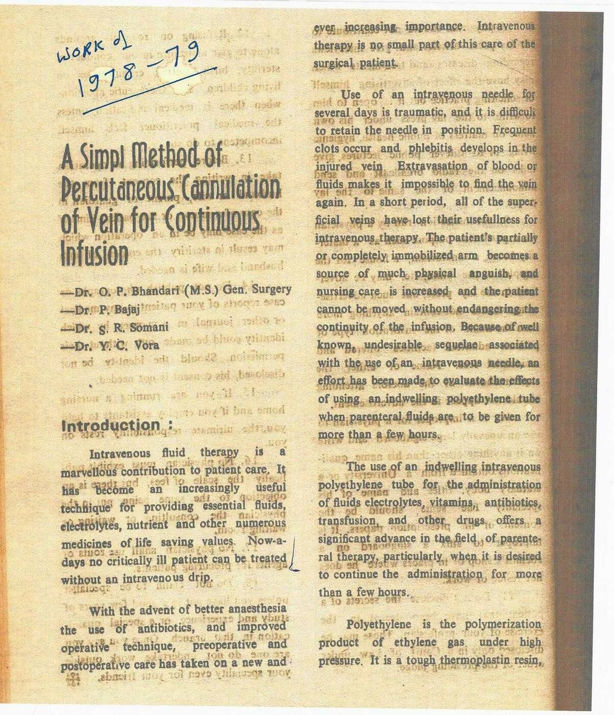
Intravenous fluid therapy is a marvellous contribution to patient care. It has become an increasingly useful technique for providing essential fluids, electrolytes, nutrients and other numerous medicines of life-saving values. Nowadays, no critically ill patient can be treated without an intravenous drip.
With the advent of better anaesthesia and the use of antibiotics, and improved operative technique, preoperative and postoperative care has taken on a new and ever-increasing importance. Intravenous therapy is no small part of this care of the surgical patient.
Use of an intravenous needle for several days is traumatic, and it is difficult to retain the needle in position. Frequent clots occur and phlebitis develops in the injured vein. Extravasation of blood or fluids makes it impossible to find the vein again. In a short period, all of the superficial veins have lost their usefulness for intravenous therapy. The patient’s partially or completely immobilised arm becomes a source of much physical anguish, and nursing care is increased and the patient cannot be moved without endangering the continuity of the infusion. Because of these well known, undesirable sequelae associated with the use of an intravenous needle, an effort has been made to evaluate the effects of using an indwelling polyethylene tube when parenteral fluids are to be given for more than a few hours.
The use of an indwelling intravenous polyethylene tube for the administration of fluids, electrolytes, vitamins, antibiotics, transfusion, and other drugs offers a significant advance in the field of parenteral therapy, particularly when it is desired to continue the administration for more than a few hours.
Polyethylene is the polymerization product of ethylene gas under high pressure. It is a tough thermoplastic resin, resistant to many chemicals. It can be sterilised by autoclaving and can be manufactured in very small diameter tubes.
II. The Innovation — Method and Materials
During the period 1977–1979, while posted to the tetanus ward of Ravindra Nath Tagore Medical College, Udaipur as a second-year postgraduate student in General Surgery, the senior author (O.P. Bhandari) encountered a recurring and serious clinical problem: tetanus patients requiring prolonged intravenous therapy rapidly exhausted all available superficial veins due to repeated needle punctures, phlebitis, and extravasation.
In the absence of commercially available intravenous cannulae — which were not yet accessible in this resource-limited district-level setting — a simple method was devised using materials that were available:
The Technique — As Originally Devised (1977–79)
Materials used: A standard BT (Blood Transfusion) set needle was used as the introducer. A length of venesection tubing — fine-bore polyethylene tubing — served as the indwelling catheter.
Procedure: The BT set needle was introduced percutaneously into the selected vein. The polyethylene tubing was then threaded through the needle lumen into the vein and advanced to the desired length. The needle was withdrawn over the tubing, leaving the catheter in situ within the vein lumen. The external end of the tubing was connected to the infusion set.
Principle: This method independently derived the principle of percutaneous venous cannulation — subsequently recognised internationally as the “over-the-needle” or “through-the-needle” catheter technique and described in the Seldinger wire-guided cannulation literature — without prior knowledge of the published international technique.
III. Clinical Setting and Context
The tetanus ward of a government medical college in Rajasthan in the late 1970s presented conditions of extreme clinical demand and minimal resources. Tetanus patients — admitted in varying stages of trismus, opisthotonus, and autonomic instability — required prolonged intravenous therapy lasting many days or weeks. Standard needle-based intravenous access invariably failed within 24–48 hours due to thrombosis, phlebitis, or dislodgement during muscular spasms. Venous cutdown (venesection) was the established alternative, but it was invasive, consumed venous sites permanently, and carried risks of wound infection and scarring in patients already immunocompromised.
The indwelling polyethylene catheter method described here provided a solution that allowed intravenous access to be maintained for several days at a single site, with the catheter held in the vein by the hydrostatic pressure of the infusion and a simple adhesive dressing. The technique was applied repeatedly during the 1977–79 period and found to be reliable, safe, and reproducible under the conditions of the tetanus ward.
IV. Significance — A 2026 Commentary
The Seldinger technique of wire-guided vascular access was first described by Sven-Ivar Seldinger in 1953 in Acta Radiologica. Commercial over-the-needle intravenous cannulae (including the Venflon and similar devices) began to become available internationally in the 1960s, and in India progressively through the 1970s and 1980s. However, in district and government medical college settings in Rajasthan in the late 1970s, such devices were not yet routinely available.
The significance of the work described in this paper lies not in claiming priority over the Seldinger or over-the-needle techniques — it does not. It lies instead in the independent derivation of the same fundamental principle by a second-year postgraduate student confronting a clinical emergency with whatever was at hand. This is precisely the nature of necessity-driven innovation: it does not require prior knowledge of the international literature; it requires only a clear understanding of the clinical problem, a disciplined mind, and the materials available.
This paper is reproduced here as a historical document because it illustrates a principle that is central to the founding conviction of the Global Science Portal for Surgical, Procedural and Clinical Innovations: that innovation belongs to every surgeon, engineer, and scientist who confronts necessity with discipline, regardless of the institutional infrastructure available to them. The tetanus ward of RNT Medical College, Udaipur, in 1977–78 was that surgeon’s laboratory.
V. The Archival Document
The original paper, annotated in the senior author’s hand as “Work of 1978–79”, is reproduced below. It was written and submitted during the postgraduate training period and represents the first formal documentation of this technique as practised at RNT Medical College, Udaipur.
VI. Authors
| Dr. O. P. Bhandari | MS (General Surgery), Ravindra Nath Tagore Medical College, Udaipur (1980). Currently: Consultant Surgeon & Director, Dr. Bhandari Piles Hospital, Chittorgarh; Founding Editor-in-Chief, GSP-SPCI; Founder General Secretary, Global Scientist Forum (est. 2000). MRSH (London), FAIS. ORCID: 0009-0008-3982-0950. |
| Dr. P. Bajaj | Department of Anaesthesiology, Ravindra Nath Tagore Medical College, Udaipur (1977–79). |
| Dr. S. R. Somani | Department of Anaesthesiology, Ravindra Nath Tagore Medical College, Udaipur (1977–79). |
| Dr. Y. C. Vora | Professor & Head, Department of Anaesthesiology, Ravindra Nath Tagore Medical College, Udaipur (1977–79). |
- Nature of Publication
- Historical paper — original work performed 1977–1979; reproduced and contextualised in GSP-SPCI 2026 with commentary by the senior author.
- Conflict of Interest
- None declared.
- Funding
- No external funding. Work performed during routine postgraduate clinical duties at RNT Medical College, Udaipur.
- Ethics
- This paper describes a clinical technique applied in routine patient care during the postgraduate training period. No formal ethics approval was required at the time of original work (1977–79). The technique is standard clinical practice today.
- Correspondence
- Dr. Om Prakash Bhandari, MS, MRSH (London), FAIS · Dr. Bhandari Piles Hospital, 4-5 Shastri Nagar, Chittorgarh – 312001, Rajasthan, India · opbhandari@hotmail.com · WhatsApp: +91-9414109124 · ORCID: 0009-0008-3982-0950