Background
Spillage of gallstones during laparoscopic cholecystectomy is a well-recognised event, reported in 6–30% of cases, and retained stones are an established cause of delayed port-site sinus and abdominal-wall abscess.(1,2,4) Loss of an appendicolith at the retrieval port during laparoscopic appendicectomy is far less often described, and its potential to mimic a controlled enterocutaneous fistula (ECF) has, to the best of a literature search performed by the author, not previously been reported.
Case Presentation
A 22-year-old man presented one month after laparoscopic appendicectomy performed elsewhere, with high-grade fever and copious, foul-smelling, faeculent discharge from the umbilical port site. The abdomen was soft and non-tender, with no peritonitis, and he continued to tolerate oral feeds and pass flatus and stool normally. A presumptive diagnosis of controlled ECF was made, and he was managed conservatively with antibiotics and regular dressings for approximately one week without improvement. During a dressing change, three variably sized calculi — later identified as appendicoliths embedded in the port tract at the time of specimen retrieval — were palpated and extracted from the wound. Discharge ceased immediately, and the sinus healed completely and rapidly thereafter.
Conclusion
Appendicoliths inadvertently implanted in the abdominal wall during unprotected specimen extraction can act, like spilled gallstones, as a nidus for chronic wound sepsis and can convincingly mimic an enterocutaneous fistula. Digital or instrumental interrogation of a non-healing port-site sinus for a retained calculus should be considered before a diagnosis of true fistula is accepted, and specimen retrieval — particularly of a friable or perforated appendix — should routinely employ a retrieval bag or ligation of the appendicular lumen to prevent this avoidable complication.
Laparoscopic appendicectomy is now an established treatment for acute appendicitis. Appendicoliths are not rare findings in this setting: on cross-sectional imaging they are noted incidentally in roughly 2.6% of scans performed for abdominal pain, and are identified in a proportion of surgically excised appendices, particularly in gangrenous or perforated appendicitis.(11) Spillage of the contents of an inflamed or gangrenous appendix — including an appendicolith — both into the peritoneal cavity and at the retrieval port during extraction of the specimen is therefore not uncommon. The analogous problem of gallstone spillage during laparoscopic cholecystectomy is well documented, occurring in 6–30% of cases,(1,2) and retained stones are known to behave as a chronic nidus for infection, producing intra-abdominal abscess, persistent abdominal-wall sinus tracts, and even distant migration, at a mean interval of roughly 27 weeks after the index operation.(4) A comparable mechanism has been reported for appendicoliths spilled or retained within the peritoneal cavity during appendicectomy, presenting months later as a subphrenic or intraperitoneal abscess.(5,6) What has not, to the author's knowledge, been previously described is a stone from the appendix becoming embedded within the port tract itself — rather than the peritoneal cavity — at the moment of specimen retrieval, and then behaving clinically as a controlled faecal fistula. This report describes such a case and reviews the mechanism by which it is thought to occur.
Mr 'X' was a 22-year-old man from an agrarian background in rural Rajasthan, who presented one month after undergoing laparoscopic appendicectomy at another facility. Despite specific effort, records of that index surgery — the operative note, including any per-operative pathological description such as gangrenous change, and the discharge summary — could not be produced by the patient or his family at the time of presentation and could not subsequently be traced. No record of the method or port used for specimen retrieval at the index operation was available either. This is a recognised practical limitation of case documentation in resource-limited, rural referral practice, where patients often present between institutions without a portable or retrievable paper trail, and it is stated here explicitly rather than left unacknowledged. Notably, at no stage — neither clinically at the index admission nor on any imaging, including the ultrasonography report the patient carried with him, which documented only a non-specifically inflamed appendix — had an appendicolith been diagnosed; no CT or MRI had been performed. Consequently, the exact operative findings at the index procedure (whether the appendix was gangrenous or perforated, whether an appendicolith was noted intra-operatively, and the precise method of specimen extraction) are not known with certainty. In the absence of any operative record, it was assumed — rather than confirmed — that specimen retrieval had been carried out through the umbilical port without a retrieval bag; this assumption, and the reasoning behind it, is discussed further below. No relevant comorbidity was elicited on history at presentation.
He presented with high-grade fever and copious, foul-smelling, dirty faecal-like discharge from the umbilical port. On examination, the abdomen was soft, with no signs of peritonentis or distension, and there was no systemic sepsis requiring admission. He continued to tolerate oral feeds and to pass flatus and stool regularly, arguing against a true high-output enteric fistula. Detailed vital-sign readings and formal wound measurements from this outpatient encounter were not preserved in the clinical record and cannot be reconstructed after the fact; this is noted as a limitation rather than presented as data that do not exist.
Abdominal ultrasonography showed no evidence of any intra-abdominal collection or fluid tracking to suggest a deep-seated source for the discharge. The combination of copious, feculent discharge from the umbilical port together with clinical signs suggestive of intraperitoneal sepsis, in the absence of a demonstrable bowel communication, led to the working diagnosis of a controlled enterocutaneous fistula following laparoscopic appendicectomy, and further, more invasive cross-sectional imaging was planned to confirm this. Before that investigation could be arranged, however, stone-like objects were incidentally palpated and retrieved during a routine outpatient dressing change, and the sinus healed spontaneously within about a week of their removal — an outcome that was taken, in the absence of formal confirmatory testing, as sufficient in itself to establish the retrospective diagnosis of an implanted appendicolith rather than a true fistula. Formal laboratory investigation (leucocyte count, inflammatory markers) and wound-swab culture were not obtained as part of this patient's outpatient-based management, and no cross-sectional imaging beyond the initial ultrasonography was in fact performed, since it was overtaken by this incidental finding; these represent limitations of the available workup rather than negative findings, and are acknowledged as such.
A persistently discharging port-site sinus after appendicectomy raises several possibilities besides a true enteric fistula: a retained surgical foreign body (suture, mesh, or gauze fragment); a stitch or ligature granuloma; port-site actinomycosis or atypical mycobacterial infection; and, less likely at this interval, a missed Crohn's-related enterocutaneous fistula. True enterocutaneous fistula is itself a recognised, if uncommon, complication of complicated laparoscopic appendicectomy — reported in around 2% of cases with complicated appendicitis, typically following percutaneous drainage of an intra-abdominal collection(12) — which is precisely why it was a reasonable initial working diagnosis here, and why its exclusion needed to be made explicit rather than assumed. The eventual extraction of calculi confirmed the diagnosis of an implanted appendicolith acting as a foreign-body nidus, but this differential should be stated explicitly for the reader's benefit, since most of these alternatives are managed very differently.
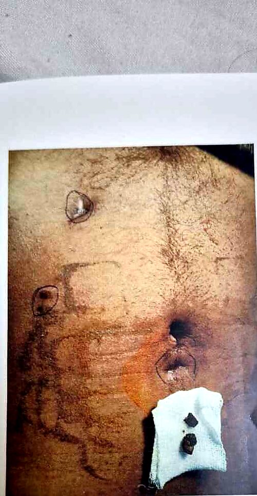
He was initially managed on empirical antibiotics, antipyretics, and regular dressings, in the expectation that a controlled fistula would close spontaneously. There was little or no improvement over approximately one week. During a subsequent dressing change, a hard, stone-like object was felt to click against the probing forceps; with gentle traction, three variably sized calculi were extracted from the sinus tract (Figure 1). Discharge ceased immediately thereafter, and the wound healed rapidly and completely within a few days (Figure 2), without further intervention.
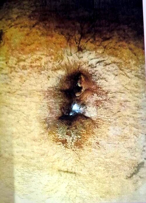
On gross inspection, the three stones were dark brown in colour with a laminated cut/surface appearance, consistent with the typical appearance of appendicoliths. Formal measurement and biochemical, histopathological, or mineral-composition analysis of the extracted stones were not performed. The identification of the calculi as appendicoliths therefore rests on their gross appearance (Figure 1), the clinical context of a recent appendicectomy, and the abrupt, complete resolution of the sinus once they were removed, rather than on laboratory confirmation. As Cruz-Santiago et al. have shown, a calculus found in or near the appendix is not invariably an appendicolith on visual inspection alone, and in principle a retained gallstone or other calcified foreign body cannot be entirely excluded on the evidence available.(13) This is stated explicitly as the principal evidentiary limitation of this report.
| Time point | Event |
|---|---|
| Day 0 | Laparoscopic appendicectomy performed elsewhere; specimen retrieved through umbilical port. (Operative record not available — see Limitations.) |
| ~4 weeks | Presentation with high-grade fever and foul-smelling faeculent discharge from the umbilical port; managed elsewhere without benefit before referral. |
| Presentation to author | Diagnosed presumptively as controlled enterocutaneous fistula; started on antibiotics, antipyretics, and regular dressing. |
| ~1 week later | Minimal to no improvement in discharge despite conservative management. |
| Index dressing change | Stone-like object palpated; three appendicoliths extracted from sinus tract. |
| Following days | Discharge ceased immediately; sinus healed rapidly and completely. |
| Follow-up | Wound confirmed completely healed (Figure 2); patient did not return for any further follow-up visit thereafter, so no later information is available. |
Spontaneous and rapid healing of the wound followed removal of the stones, allowing the retrospective diagnosis of a spontaneously implanted appendicolith at the port site mimicking an enterocutaneous fistula. Once the wound had healed completely, the patient did not return for any further follow-up visit — a common pattern in this rural referral setting once the presenting complaint has resolved. Consequently, there is no documented information, one way or the other, on recurrence, incisional hernia, or late discharge beyond the point of confirmed complete healing; this should be read as an absence of follow-up data rather than as confirmed absence of any such complication.
Port-site stitch infection, subcutaneous or superficial abscess, wound dehiscence, and incisional hernia are frequently documented complications of laparoscopic cholecystectomy,(3) and a comparable spectrum of complications is recognised after laparoscopic appendicectomy. Retained or spilled gallstones are known to provoke adhesions and a persistent inflammatory reaction without meaningful absorption, and can present, on average, some 27 weeks after the index surgery as an intra-abdominal abscess, an abdominal-wall sinus tract, or even, rarely, a stone that has migrated to a joint space.(4) A directly analogous process has been reported for appendicoliths retained within the peritoneal cavity, which have subsequently presented as a subphrenic collection(5) or required image-guided localisation for surgical retrieval once symptomatic.(6) These reports establish that both gallstones and appendicoliths, once retained in a non-physiological space, behave as inert-appearing but biologically active foreign bodies capable of sustaining chronic low-grade sepsis.
The present case differs from those reports in an important respect: the stones were not lost into the peritoneal cavity, but became mechanically embedded within the substance of the port tract itself, presumably at the moment the appendix — possibly gangrenous or perforated and therefore friable, though this cannot be confirmed in the absence of the index operative record — was withdrawn through an unprotected port without a retrieval bag. A calcified appendicolith caught in the fascial or subcutaneous layers during extraction would be expected to behave exactly as it did here: a discharging sinus that persists despite antibiotics because the nidus (the stone) has not been removed, that fails to communicate with bowel on imaging because there is no true fistula, and that heals abruptly and completely the moment the foreign body is extracted. This mechanism, and the resulting clinical picture of a "pseudo-fistula," is a useful addition to the differential diagnosis of a non-healing port-site sinus, alongside stitch granuloma, retained gauze, and port-site actinomycosis.
A search of PubMed/MEDLINE (search terms: "appendicolith," "port site," "implanted," "enterocutaneous fistula," "laparoscopic appendicectomy," undertaken at the time of this revision) did not identify a previously published case of an appendicolith implanted specifically at a laparoscopic port site producing this clinical picture; the closest precedents concern appendicoliths spilled into the peritoneal cavity rather than caught within the abdominal wall itself.(5,6) This report should therefore be read as an apparently rare, and possibly novel, presentation rather than as definitive proof of a "first-ever" case, since the possibility of unpublished or non-indexed prior reports cannot be excluded.
This report has four principal limitations, stated here explicitly rather than left implicit. First, it describes a single patient, and the mechanism proposed for stone implantation at the port tract is inferred from the clinical course rather than observed directly. Second, records of the index laparoscopic appendicectomy performed at another facility could not be obtained despite specific effort — a genuine constraint of caring for patients from underserved rural backgrounds who present between institutions without a portable clinical record — so the operative findings and method of specimen extraction at the first surgery, including the assumption that retrieval was through the umbilical port without a bag, are inferred rather than documented. Third, the three extracted calculi were not sent for biochemical, histopathological, or mineral-composition analysis, so their identification as appendicoliths rests on gross appearance and clinical context rather than laboratory confirmation, and cannot be regarded as definitive. Fourth, the patient did not return for any follow-up visit once the wound had healed, so the medium- and long-term outcome — including whether any recurrence or incisional complication occurred — is simply not known rather than confirmed to be absent. None of these limitations alter the clinical fact around which this report is built — that discharge from a non-healing port-site sinus, mimicking an enterocutaneous fistula, resolved immediately and completely upon extraction of implanted calculi — but they should be borne in mind by the reader.
This mechanism has a direct bearing on operative practice. Several retrospective series report lower surgical-site infection rates when the specimen is protected from direct contact with the port wound during extraction — whether by a purpose-made retrieval bag, or by the low-cost expedient of a sterile surgical glove used as an improvised bag or "finger cot."(8,9,10) However, this is not entirely settled: a randomised controlled trial comparing bagged versus unprotected extraction in 165 laparoscopic appendicectomies found no significant difference in surgical-site infection between the two groups, and concluded that a retrieval bag does not by itself prevent microbial seeding of the wound.(9) Taken together, the literature supports protecting the port wound from direct specimen contact as good practice, but does not establish it as sufficient on its own to eliminate wound complications — mechanical loss of a hard, faceted appendicolith against the fascia during extraction, as is proposed here, is a distinct problem from bacterial contamination and may not be fully addressed by a retrieval bag unless the bag itself is handled carefully during withdrawal.
A cautionary parallel from the literature is worth noting: a stone found at the base of the appendix and presumed on imaging to be an appendicolith has, in at least one reported case, proved on histopathological analysis to be an impacted gallstone that had migrated via a choledocho-duodenal fistula and caused an obstructive gangrenous appendicitis.(13) That case is a useful reminder that a calculus found in or around the appendix is not invariably a true appendicolith on visual inspection alone, and it reinforces — rather than merely as an editorial preference — why sending the three stones recovered in the present case for histopathological or mineralogical analysis would materially strengthen the diagnosis, rather than resting on gross appearance and clinical context alone.
Take-Home Messages
- A non-healing, foul-smelling port-site sinus after laparoscopic appendicectomy that fails to respond to antibiotics should prompt digital or instrumental probing for a retained calculus before a diagnosis of true enterocutaneous fistula is accepted.
- An implanted appendicolith can produce a clinical picture that is difficult to distinguish from a controlled enteric fistula, yet resolves abruptly and completely once the foreign body is removed — a pattern that is itself diagnostic in retrospect.
- To prevent spillage of appendicoliths and other appendiceal contents during specimen retrieval, the cut end of the appendicular lumen should be ligated before extraction.
- A gangrenous or bulky appendix that cannot be delivered comfortably through a 10 mm or 12 mm cannula should be placed in a retrieval (endo-) bag, or at minimum a sterile glove used as an improvised bag, before extraction — good general practice for wound protection, though the evidence on whether this alone prevents every wound complication is mixed and it should not be relied on in isolation.(8,9,10)
- Intra-operative documentation should record whether an appendicolith was identified, whether it was retrieved intact, and the method of specimen extraction used — mirroring existing recommendations for documenting gallstone spillage.(4)
Port-site implantation of gallbladder stones is a well-known entity; the literature reviewed for this report did not reveal a previously published case of an appendicolith implanted specifically at a laparoscopic port site in this manner. Subject to the transparency caveats noted in the Discussion, this appears to be a rare, and possibly novel, presentation worth documenting for the awareness of surgeons managing non-healing wounds after laparoscopic appendicectomy.
Conflict of Interest
No conflict of interest.
Funding
No funding was received for this work.
Ethics / Consent
Written (or, where literacy was a barrier, witnessed verbal) informed consent for publication of clinical details and images was obtained from the patient; he is referred to as Mr. 'X' throughout so that he cannot be identified. As a single-patient retrospective case report involving no experimental intervention, this work is considered exempt from formal institutional ethics committee review under prevailing case-report reporting norms; no departure from standard clinical care occurred.
Author Contributions
N.K.M. managed the patient, conceived the report, reviewed the literature, and wrote and approved the final manuscript.
Data Availability
Clinical data supporting this report are held in the patient's medical records and are available from the corresponding author on reasonable request, subject to patient confidentiality.
Reporting Guideline
This case report has been prepared in accordance with the CARE (CAse REport) guidelines.(7)
- Diez J, Arozamena C, Gutierrez L, et al. Lost stones during laparoscopic cholecystectomy. HPB Surg. 1998;11:105-109.
- Hand AA, Self ML, Dunn E. Abdominal wall abscess formation two years after laparoscopic cholecystectomy. JSLS. 2006 Jan-Mar;10(1):105-107.
- Kumar TS, Saklani AP, Vinayagam R, Blackett RL. Spilled gallstones during laparoscopic cholecystectomy: a review of the literature. Postgrad Med J. 2004;80:77-79.
- Chin PT, Boland S, Percy JP. "Gallstone hip" and other sequelae of retained gallstones. HPB Surg. 1997;10(3):165-168. doi: 10.1155/1997/14698. PMID: 9174862.
- Whalley HJ, Remoundos DD, Webster J, Silva MA. Shortness of breath, fever and abdominal pain in a 21-year-old student. BMJ Case Rep. 2013;2013:bcr2013200729. doi: 10.1136/bcr-2013-200729. PMID: 24127375.
- Lossef SV. CT-guided Kopans hookwire placement for preoperative localization of an appendicolith. AJR Am J Roentgenol. 2005;185(1):81-83. doi: 10.2214/ajr.185.1.01850081. PMID: 15972402.
- Gagnier JJ, Kienle G, Altman DG, Moher D, Sox H, Riley D, for the CARE Group. The CARE guidelines: consensus-based clinical case reporting guideline development. Glob Adv Health Med. 2013;2(5):38-43. doi: 10.7453/gahmj.2013.008. PMID: 24416692.
- Bostan MS, Uğurlu C. Is it necessary to use a specimen retrieval bag for reducing surgical site infection in laparoscopic appendectomy? A randomized controlled trial. Ulus Travma Acil Cerrahi Derg. 2023;29(2):203-211. doi: 10.14744/tjtes.2022.97828. PMID: 36748777.
- Agalar C, Derici S, Çevlik AD, Aksoy SÖ, Egeli T, Boztaş N, Özbilgin M, Sarıoğlu S, Ünek T. Do the stump knotting technique and specimen retrieval method affect morbidity in laparoscopic appendectomy? Ulus Travma Acil Cerrahi Derg. 2019;25(1):34-38. doi: 10.5505/tjtes.2018.90382. PMID: 30742284.
- Aytac HO, Torer N, Arer IM. The type of specimen retrieval in laparoscopic appendectomy affects wound infection. Ann Ital Chir. 2016;87:572-576. PMID: 28070028.
- Islam S, Bheem V, Maughn A, Harnarayan P, Dan D, Naraynsingh V. Surgical glove use for specimen removal in laparoscopy, the cheapest available: a prospective study. Trop Doct. 2019;50(1):94-99. doi: 10.1177/0049475519871861. PMID: 31495274.
- Rollins MD, Andolsek W, Scaife ER, Meyers RL, Duke TH, Lilyquist M, Barnhart DC. Prophylactic appendectomy: unnecessary in children with incidental appendicoliths detected by computed tomographic scan. J Pediatr Surg. 2010;45(12):2377-2380. doi: 10.1016/j.jpedsurg.2010.08.038. PMID: 21129549.
- Khiria LS, Ardhnari R, Mohan N, Kumar P, Nambiar R. Laparoscopic appendicectomy for complicated appendicitis: is it safe and justified? A retrospective analysis. Surg Laparosc Endosc Percutan Tech. 2011;21(3):142-145. doi: 10.1097/SLE.0b013e31821ad770. PMID: 21654295.
- Cruz-Santiago J, Briceño-Sáenz G, García-Álvarez J, Beristain-Hernández JL. Gallstone ileus presenting as obstructive gangrenous appendicitis. Rev Esp Enferm Dig. 2017;109(2):150-151. PMID: 28211283.
Professor of Surgery, Ex-HOD, SMS Medical College, Jaipur, and RNT Medical College, Udaipur
Director & Chief Surgeon, Malpani Multispeciality Hospital, V.K.I.A., Sikar Road, Jaipur, Rajasthan, India Address 7/67, New Vidhyadhar Nagar, Jaipur – 302039, Rajasthan, India Email [to be supplied — journal metadata requires a corresponding-author email] Peer Reviewer Dr. Sandeep Purohit, MCh (Urology), RNT Medical College, Udaipur — reviewed and cleared for publication